Call Us: (267) 727-3738

MRI in Veterinary Practice

Magnetic resonance imaging (MRI) is an advanced, noninvasive diagnostic imaging modality that has become an invaluable resource in veterinary specialty practice. Its superior soft-tissue contrast makes MRI particularly useful for neurologic and musculoskeletal cases where radiographs, ultrasound, or computed tomography (CT) may be inconclusive or unrewarding. MRI is most commonly available in specialty and academic settings, but has become increasingly available.
At PASE, our 1.5T high-field MRI is utilized by the Neurology and Neurosurgery, Diagnostic Imaging, and Surgery teams, allowing us to diagnose both routine and complex conditions.
What Is MRI?
MRI uses a powerful magnetic field and radiofrequency (RF) waves to create detailed images of the body without ionizing radiation. When a patient is placed in the magnetic field, hydrogen protons within tissues align with that field. RF pulses temporarily disrupt this alignment, and as the protons relax back to baseline, they emit signals that are detected and processed into images. Differences in tissue composition—such as water content, fat, and molecular environment—produce variations in signal intensity, allowing excellent differentiation between tissues.
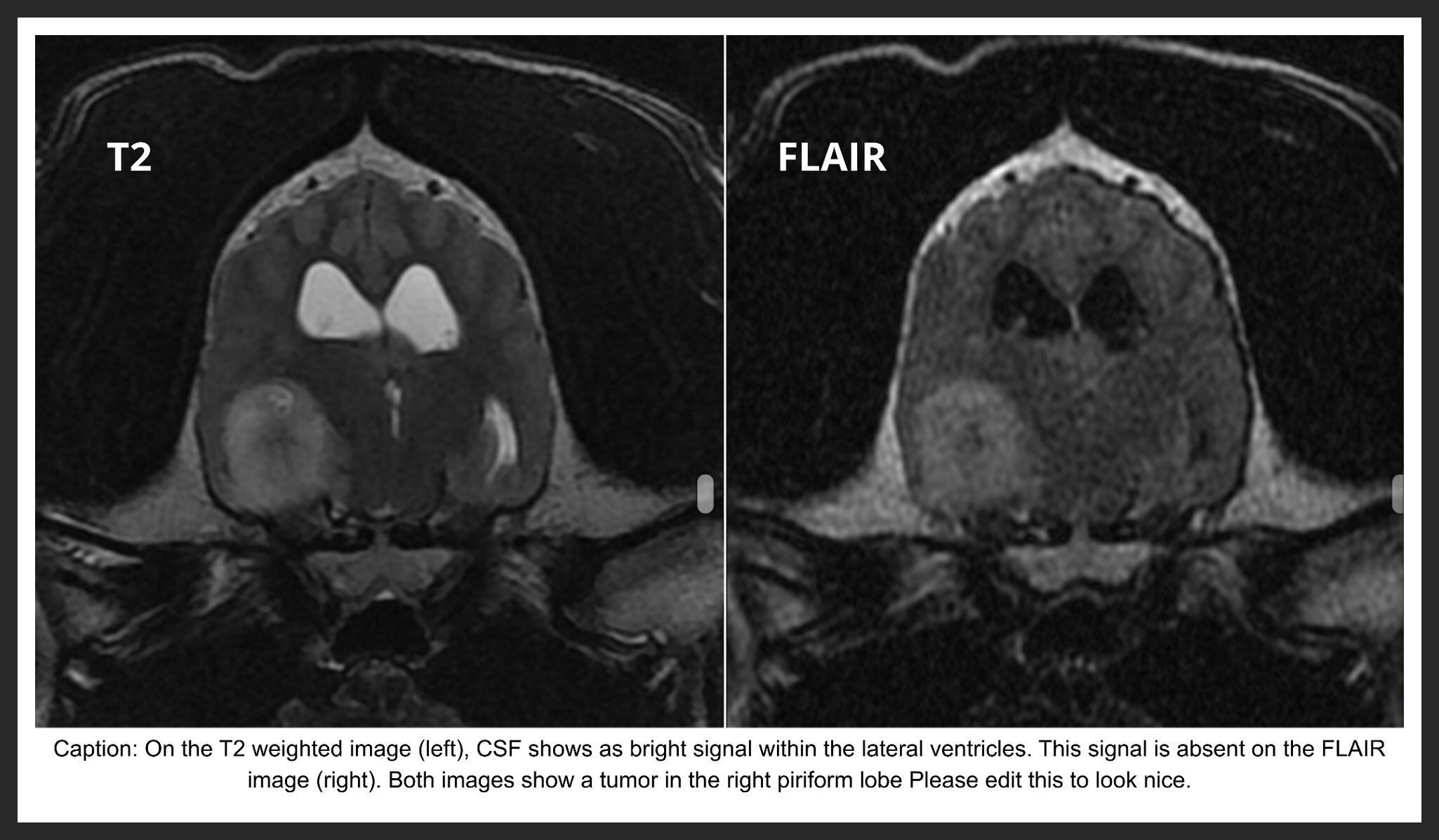
RF pulses are applied in various imaging “sequences,” each designed to highlight specific physical or chemical tissue characteristics. These sequences are interpreted together to build a comprehensive diagnostic picture. For example, a FLAIR (fluid-attenuated inversion recovery) sequence suppress signal from free fluid and is routinely used in neuroimaging to eliminate cerebrospinal fluid signal, improving evaluation of periventricular brain tissue and highlighting edema. Other sequences suppress fat signal (e.g., STIR, SPAIR, spectral fat saturation) to accentuate inflammation or fluid accumulation. Additional specialized sequences can also detect hemorrhage. As with CT, contrast agents may be administered to further characterize inflammation, vascularity, or neoplasia.
High-field MRI systems (1.5T or greater) provide higher image resolution, improved soft-tissue contrast, and shorter scan times compared to low-field systems. This enhanced image quality allows better detection of subtle lesions, particularly in neurologic and complex musculoskeletal cases. While low-field MRI can be useful in select situations, high-field MRI offers the most comprehensive diagnostic information.
When Is MRI Indicated?
Neurologic Applications
High-field MRI is the gold standard imaging modality for the brain and spinal cord. Direct visualization of neural tissue allows accurate diagnosis, treatment planning, and prognostication. Common indications include:
- Intervertebral disc disease
- Spinal cord compressive disorders other than IVDD
- CNS vascular injury or ischemic events
- Brain, spinal cord, or peripheral nerve sheath tumors
- Seizure disorders, particularly with suspected structural disease
- Inflammatory or infectious CNS disease
- Congenital abnormalities
- Vertebral or paravertebral masses
- Lumbosacral disease
Musculoskeletal Applications
MRI is increasingly used to evaluate soft-tissue orthopedic disease, especially when other imaging modalities fail to identify a cause. Indications include:
- Joint disease, particularly of the shoulder and stifle
- Tendon and ligament injuries
- Muscle injuries
- Cartilage defects
- Unexplained or persistent lameness
- Musculoskeletal neoplasia
Referral Information
If you have a case that may benefit from MRI evaluation, please feel free to contact our Neurology or Surgery teams for additional information or to discuss referral options.
Referrals can be submitted directly through our portal at www.pase.vet/referral-portal.html, or you may call 267-727-3738 to make an appointment.
PASE’s Neurology service sees appointments Monday through Saturday and is on call every Sunday.
PASE’s Surgery service sees appointments Monday through Friday.
Phone
Hours
24 Hours A Day / 7 Days A Week
Affiliate Hospital
NorthStar VETS
(609) 259-8300
3 New Jersey Locations
northstarvets.com
© 2026 Philadelphia Animal Specialty & Emergency. All rights Reserved.
Privacy Policy - Sitemap
Powered by